Back أديم متوسط Arabic Mezoderma Azerbaijani Мезодерма Bulgarian Mezoderm BS Mesoderma Catalan Mezoderm Czech Mesoderm Danish Mesoderm German Μεσόδερμα Greek Mezodermo Esperanto
This article needs additional citations for verification. (May 2022) |
| Mesoderm | |
|---|---|
 Tissues derived from mesoderm. | |
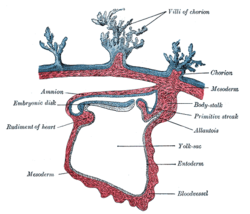
 Section through a human embryo | |
| Details | |
| Days | 16 |
| Identifiers | |
| MeSH | D008648 |
| FMA | 69072 |
| Anatomical terminology | |
The mesoderm is the middle layer of the three germ layers that develops during gastrulation in the very early development of the embryo of most animals. The outer layer is the ectoderm, and the inner layer is the endoderm.[1][2]
The mesoderm forms mesenchyme, mesothelium, non-epithelial blood cells and coelomocytes. Mesothelium lines coeloms. Mesoderm forms the muscles in a process known as myogenesis, septa (cross-wise partitions) and mesenteries (length-wise partitions); and forms part of the gonads (the rest being the gametes).[1][unreliable source?] Myogenesis is specifically a function of mesenchyme.
The mesoderm differentiates from the rest of the embryo through intercellular signaling, after which the mesoderm is polarized by an organizing center.[3] The position of the organizing center is in turn determined by the regions in which beta-catenin is protected from degradation by GSK-3. Beta-catenin acts as a co-factor that alters the activity of the transcription factor tcf-3 from repressing to activating, which initiates the synthesis of gene products critical for mesoderm differentiation and gastrulation. Furthermore, mesoderm has the capability to induce the growth of other structures, such as the neural plate, the precursor to the nervous system.
- ^ a b Ruppert, E.E.; Fox, R.S.; Barnes, R.D. (2004). "Introduction to Bilateria". Invertebrate Zoology (7th ed.). Brooks/Cole. pp. 217–218. ISBN 978-0-03-025982-1.
- ^ Langman's Medical Embryology, 11th edition. 2010.
- ^ Kimelman, D. & Bjornson, C. (2004). "Vertebrate Mesoderm Induction: From Frogs to Mice". In Stern, Claudio D. (ed.). Gastrulation: from cells to embryo. CSHL Press. p. 363. ISBN 978-0-87969-707-5.
© MMXXIII Rich X Search. We shall prevail. All rights reserved. Rich X Search